[ general | scan action | scaling | file | context menu || main help index ]
There are some small GUI elements which can be found in similar versions in different windows or group boxes. Those elements are shortly explained here.
A small button next to the upper right edge of some group box frames can minimize or maximize the corresponding group box. This is very helpful to make the windows as small as possible in vertical direction.
There are two time displays on the upper side of some scan settings group box frames.

The left one shows the elapsed time of the currently running scan, the right one is a rough estimate of the total scan time, i.e.,
T=Nx*Ny*tgate
Depending on your computer, the data acquisition hardware, other software running and the number of active scans, significant latency times may occur. Thus the total scan time is usually between 1% and 5% greater than the estimate.
Note: The elapsed scan time is the sum of the time slices where the scan thread was active. If multiple scans are executed, then the progress of each of them is slowed down and the elapsed time will not proceed in real time.
Note: It is generally recommended to use gate times that are multiples of 10 ms for longer scans. The program is then able to give back some CPU time to the operating system: A minimum CPU load is the result.
In many scan windows there are two buttons showing a clipboard and an arrow pointing either to or from the clipboard. You can use them to copy a set of scan parameters as a text message to or from the Windows clipboard. For example, this allows you to select a rectangular region in a 2D scan (with the rectangle tool), copy the coordinates to the clipboard and use these data for a new 2D scan. Or you can select a line in a 2D scan (with the line tool), get a 1D scan across this line, and then decide to make a reciprocal space map vertical to the original 2D scan along the 1D scan.
Here you can choose how and when the specified SPA-LEED scan should be executed.

| GUI element | Meaning and Usage |
|---|---|
| Start button | Click here to start a scan. After starting
a scan, this button can be clicked again to cancel the scan, i.e.,
to stop it immediately. Note: This is a big difference to the Spa4.1d software. If you just want to finish a repeating scan as soon as possible, don't click on cancel but change the scan mode instead. |
| Scan mode radio buttons | There are three possible scan modes available:
"stop at end", "continuous" or "repetitive".
The first one is the most usual scan mode in any scanning
microscopy; a scan is stopped
after the last scan line has been finished. In many cases, however, it is not possible to decide at the beginning of a scan which gate time (and thus: scan quality) is optimal. You might think after one third of a scan "Why didn't I use higher gate times, it's a great pattern?" or "Why the hell am I scanning this crap? It will take another two hours!" For those cases you can use the continous scan mode: The same scan is executed over an over again and the counts are accumulated (unthinkable for an STM because of sample drift!). If you choose a typical gate time of - let's say - 2 ms and a resolution of 400x400 points, a full scan will take 5 minutes. That is short enough to wait for the end of a scan if the pattern is not good, but you can let this run several times to improve the count statistics in the diffuse backgrond intensity. Finally, there is a scan mode called repetitive scan. A specified scan is executed again and again after a specified delay in between. You can use this for adjustment purposes, but in most cases this is a scan mode to observe a part of the diffraction pattern over time. In that case, you probably wish to switch on the "auto save" switch in the "file settings". Note: To stop any kind of scan after the last line has been finished, click on "stop at end", not on "Cancel". |
| delay time for repetition | A repetitive scan is usually used for long-time in-situ measurements of up to some hours where the spot(s) being observed should not be scanned all the time in order to protect the channeltron. Therefore, you can choose a delay time to - let's say - scan a specific spot only each 20 seconds. In the mean time, the electron beam is "parked", see software settings window (>DAQ>x,y beam park position). |
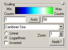
In an image scaling group box you can change the settings for the display of a 2D data set, i.e., 2D scan, SEM scan and RSM scan.

| GUI element | Meaning and Usage |
|---|---|
| Colour bar | Shows the colour table chosen with the colour table drop-down-list. |
| Min. Counts | The minimum number of counts to display, corresponding to the very left colour in the current colour table. Setting this value to more than the minimum number of counts within the scan reduces the noise in the diffuse background intensity. |
| Max. Counts | The maximum number of counts to display, corresponding to the very right colour in the current colour table. Setting this value to less than the maximum number of counts within the scan makes features close to intense spots or structures in the diffuse background intensity better visible. |
| Auto (count range) | Automatically determine the minimum and maximum number of counts within the current data set. |
| Colour table dropdown list | Here you can browse through the available colour tables. Look at the colour bar to see the chosen colour table. The change is not automatically applied to the display, since displaying a large 2D scan might take several 1-2 seconds. |
| Linear/Logarithmic | Choose whether you prefer a linear or logarithmic dependence between intensity and colour table index. In linear scaling there's usually not much more to see than the specular spot. |
| Inverted | If this is selected, the colour table is inverted (=mirrored). Try this out mainly for real space scans where an inverted colour table can inprove the visible appearance of many scans. |
| Zoom | Usually each pixel on the screen corresponds to one pixel of your scan data in order to avoid moire patterns. For scans with only few pixels it might make sense to magnify them for display. The integer number N that you enter as zoom value means that each pixel of your scan is represented by a NxN pixels sized square on your screen. |
| Apply ! | Upon clicking on this button, the (new) settings for data display are applied to the display. |
Note: In the reciprocal space map scan (RSM) you can use different zoom factors for the k||- and the kz-direction:

In WinSPA there are a couple of different colour tables available for displaying 2D scan data. Note that the colour tables with high dynamic ranges should mainly be used for scan data with good statistics, i.e., scans that were acquired with typical counter gate times of around or more than 10 milliseconds.
| Colour Table Index | Name of Colour Table | Image | Colours | Dynamic Range | Description/Comments |
|---|---|---|---|---|---|
| 0 | Black and White |
| black, white | 8 bits | The most basic colour table, grayscale between black and white. Very good for printouts and publications. |
| 1 | Fade to Red |
| black, red | 8 bits | Similar to the grayscale colour table, but turned red. |
| 2 | Fade to Green |
| black, green | 8 bits | Similar to the grayscale colour table, but
turned green. If used as inverted, it looks like a phosphor screen image ("vintage LEED"). |
| 3 | Fade to Blue |
| black, blue | 8 bits | Similar to the grayscale colour table, but turned blue. |
| 4 | Red Heat |
| black, red, white | 9 bits | ... |
| 5 | Deep Sea |
| black, blue, white | 9 bits | ... |
| 6 | Jungle |
| black, green, white | 9 bits | ... |
| 7 | Intensity |
| black, blue, green, red, white | 10 bits | This is the godfather of all false colour tables. |
| 8 | Fire |
| black, blue, red, yellow, white | 10 bits | Often used for false-colour temperature images. |
| 9 | Rainbow |
| red, yellow, green, cyan, blue | 10 bits | Not really a rainbow, but very colourful. |
| 10 | Geo |
| blue, green, yellow, red, white | 10 bits | A colour table that is similar to the standard colour table for topographical data on geography. |
| 11 | Caribbean Sea |
| black, blue, cyan, white, green | 10 bits | This colour table lets you feel like being in holiday. It shows the transition from deep water to sea, shore, beach and finally trees. |
| 12 | Interference Fringes |
| black, white | 11 bits (modulo) | This colour table is great for revealing structures in the diffuse intensity background, but it should not be used for the final data display since it does not provide a one-to-one correspondence between intensity and colour. |
| 13 | Topographic |
| black, blue, lime, green, yellow, maroon, white | 11 bits | This colour table gives you the impression of a topographic map showing deep water, sea, shore, vegetation, desert, mountains and glacier/snow. |
In an x-y scaling group box you can change the settings for the display of an x-y data set, i.e., 1D scan (left) or I(t) scan (right).

![]()
| GUI element | Meaning and Usage |
|---|---|
| Min. Cps. / Min. Cnts. | The minimum number of counts or counts per second to display. corresponding to lowest value of the y axis. |
| Max. Cps. / Max. Cnts. | The maximum number of counts or counts per second to display. corresponding to highest value of the y axis. |
| Auto cps. / cnts. | Automatically determine the minimum and maximum number of counts or counts per second within the current data set. |
| From k= / t min. | The minimum value (=left border) of the axis of ordinates (=t axis for I(t) scans, k axis for 1D scans). |
| To k= / t max. | The maximum value (=right border) of the axis of ordinates (=t axis for I(t) scans, k axis for 1D scans). |
| full range | The k-scaling is set according to the scan range (1D scan). |
| Auto t min. | The display start is automatically calculated, the displayed time span remains constant (I(t) scan). |
| Auto t max. | The display end is automatically calculated, so that the latest scan data can be displayed (I(t) scan). |
| Linear / Logarithmic | Set the scaling of the abscissa (I axis) to either linear or logarithmic scaling. |
| Apply ! | Apply the new scaling settings and display the data set. |
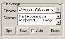
In the file settings group box you can specify all parameters about how the current data set is saved or you can load save files from your storage medium.

| GUI element | Meaning and Usage |
|---|---|
| Filename | This is the name of the current file. If the name requires more space than the width of the edit then it is truncated in the middle ("..." inserted) for easier display. Once you enter the edit the full path and name are displayed. |
| "?" = change file name/browse files | Click here to choose a file name and/or directory by browsing. |
| Comment | Your comment about the file.
You might want to add a short description of what the file contains and why you saved it, like "Si/Ge(111)@520K, satellite spots!". |
| Open | Click here to open a WinSPA or Spa4.1d data file. |
| Save | Click here to save the scan data into a WinSPA file. |
| Auto (save) | If this checkbox is activated, the scan data will
automatically be saved after the scan is finished. This is not available for the I(t) scan, since this scan does not stop automatically. The auto save setting is very important for all repetitive scans (see scan action GUIs). |
| Export | Click here to export the data as a Spa4.1d data file.
This option is available only for 1D and 2D scans. Note: This is generally not recommended, since quite some information is lost in this case. Specifically, Spa4.1d data files only handle Volts as k space units. |
The context menu appears if you right-click on any scan display. For all image-like scan types (2D, RSM, SEM, timeplot) you can export the data image without any scaling.

| GUI element | Meaning and Usage |
|---|---|
| Save as Bitmap with scaling | Saves the current scan data "as is" to a bitmap file. The image will contain exactly what you see on your screen. |
| Print out | Prints out the current scan data "as is" with the printer
specified in the software settings window. In addition, the scan parameters are also shown. Such a printout is perfectly suited for being placed in your lab book. |
| Export as ASCII | Exports the scan data in an ASCII file according to the settings for ASCII export in the File I/O tab of the software settings form. All meta-data are stored as comments. |
| Save as Bitmap (Image only) | Saves the current scan data "as is" to a bitmap file, but - in contrast to the first menu item of the context menu - without borders, axes, ticks, tickmarks etc. |